
File:Clara cell lung - TEM.jpg
No higher resolution available.
Clara_cell_lung_-_TEM.jpg (640 × 480 pixels, file size: 98 KB, MIME type: image/jpeg)
| This is a file from the Wikimedia Commons. Information from its description page there is shown below. Commons is a freely licensed media file repository. You can help. |
Summary
| Description |
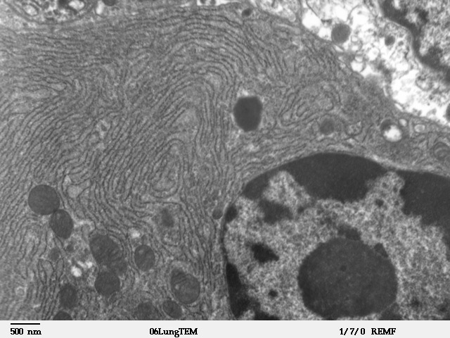
Transmission electron microscope image of a thin section cut through an area of mammalian lung tissue. This image of a bronchiolar exocrine cell (Club cell) shows a nucleus and cytoplasmic organelles, such as rough endoplasmic reticulum and mitochondria. JEOL 100CX TEM |
| Source | |
| Author | Louisa Howard |
| Permission (Reusing this file) |
PD |
Licensing
| This work has been released into the public domain by its author, Louisa Howard. This applies worldwide. In some countries this may not be legally possible; if so: Louisa Howard grants anyone the right to use this work for any purpose, without any conditions, unless such conditions are required by law.
|
File history
Click on a date/time to view the file as it appeared at that time.
| Date/Time | Thumbnail | Dimensions | User | Comment | |
|---|---|---|---|---|---|
| current | 20:39, 4 October 2006 | | 640 × 480 (98 KB) | Patho | {{Information |Description=Transmission electron microscope image of a thin section cut through an area of mammalian lung tissue. This image of a Clara cell shows a nucleus and cytoplasmic organelles, such as rough endoplasmic reticulum and mitochondria. |
File usage
The following page uses this file:
Global file usage
The following other wikis use this file:
- Usage on ar.wikipedia.org
- Usage on ckb.wikipedia.org
- Usage on cs.wikipedia.org
- Usage on de.wikipedia.org
- Usage on de.wikibooks.org
- Usage on el.wikipedia.org
- Usage on en.wikipedia.org
- Usage on eo.wikipedia.org
- Usage on fa.wikipedia.org
- Usage on gl.wikipedia.org
- Usage on gv.wikipedia.org
- Usage on ht.wikipedia.org
- Usage on hu.wikipedia.org
- Usage on hy.wikipedia.org
- Usage on jv.wikipedia.org
- Usage on kn.wikipedia.org
- Usage on mk.wikipedia.org
- Usage on ml.wikipedia.org
- Usage on mn.wikipedia.org
- Usage on ms.wikipedia.org
- Usage on pl.wikibooks.org
- Usage on ru.wikibooks.org
- Usage on sh.wikipedia.org
- Usage on si.wikipedia.org
- Usage on sl.wikipedia.org
- Usage on sr.wikipedia.org
- Usage on ta.wikipedia.org
- Usage on th.wikipedia.org
- Usage on tl.wikipedia.org
- Usage on uk.wikipedia.org
- Usage on ur.wikipedia.org