
File:Plant cell types.svg

Size of this PNG preview of this SVG file: 800 × 564 pixels. Other resolutions: 320 × 226 pixels | 640 × 451 pixels | 1,024 × 722 pixels | 1,280 × 903 pixels | 2,560 × 1,805 pixels | 1,900 × 1,340 pixels.
Original file (SVG file, nominally 1,900 × 1,340 pixels, file size: 7.16 MB)
| This is a file from the Wikimedia Commons. Information from its description page there is shown below. Commons is a freely licensed media file repository. You can help. |
Summary
| Description |
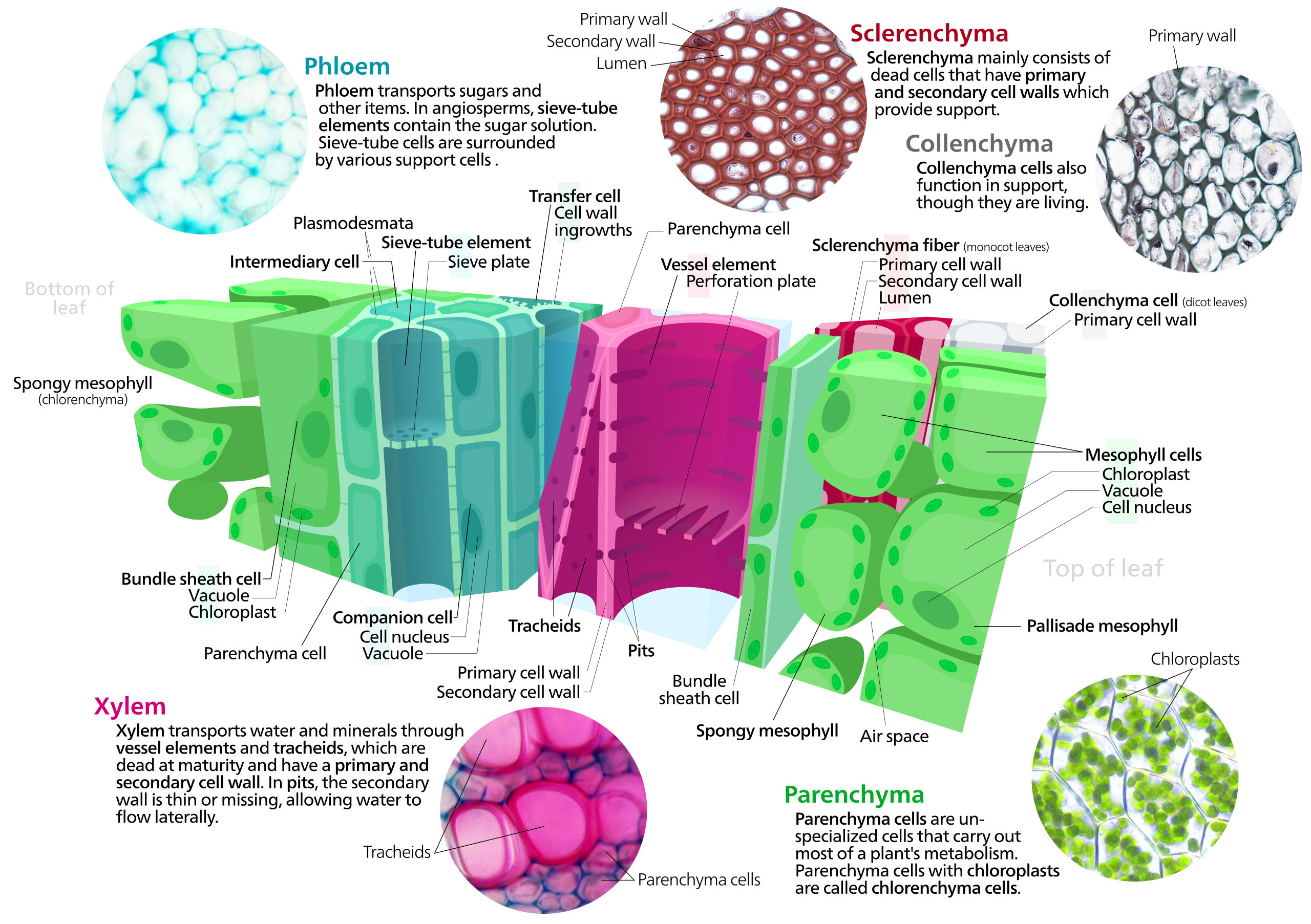
English: A cross section of a leaf showing the phloem, xylem, sclerenchyma and collenchyma, and mesophyll.
—Microscope images used— Xylem and phloem File:Lamium sp., stalk, Etzold green 5.jpg, by Micropix. Sclerenchyma and collenchyma File:Plant cell type sclerenchyma fibers.png and File:Plant cell type collenchyma.png, by Snowman frosty Chlorenchyma File:Plagiomnium affine laminazellen.jpeg by Fabelfroh —Sources— Biology—Campbell&Reece 8th edition (pgs 744, 745, 751) |
|||||||||||||||
| Date | ||||||||||||||||
| Source | Own work | |||||||||||||||
| Author | Kelvinsong | |||||||||||||||
| Other versions |
|
|||||||||||||||


|
This image was selected as picture of the day on Wikimedia Commons for 27 July 2013. It was captioned as follows: English: A cross section of a leaf showing the phloem, xylem, sclerenchyma and collenchyma, and mesophyll. Other languages:
English: A cross section of a leaf showing the phloem, xylem, sclerenchyma and collenchyma, and mesophyll. Italiano: Diagramma trasversale di una foglia che mostra il floema, xilema, sclerenchima, collenchima e mesofillo. Magyar: Egy levél keresztmetszetén ábrázolva a floém, xilém, a szklerenchima és kollenchima, valamint a mezofillum. Nederlands: Een dwarsdoorsnede van een blad toont het floëem, xyleem, sklerenchym, collenchym en mesofyl. |
Licensing
I, the copyright holder of this work, hereby publish it under the following license:
This file is licensed under the Creative Commons Attribution-Share Alike 3.0 Unported license.
- You are free:
- to share – to copy, distribute and transmit the work
- to remix – to adapt the work
- Under the following conditions:
- attribution – You must give appropriate credit, provide a link to the license, and indicate if changes were made. You may do so in any reasonable manner, but not in any way that suggests the licensor endorses you or your use.
- share alike – If you remix, transform, or build upon the material, you must distribute your contributions under the same or compatible license as the original.
File history
Click on a date/time to view the file as it appeared at that time.
| Date/Time | Thumbnail | Dimensions | User | Comment | |
|---|---|---|---|---|---|
| current | 23:11, 15 April 2013 | | 1,900 × 1,340 (7.16 MB) | IsadoraofIbiza | cut and moved text, added leaf side labels, photo labels |
| 00:03, 15 April 2013 |  | 2,000 × 1,360 (7.24 MB) | IsadoraofIbiza | fixing clipped chloroplast | |
| 20:44, 14 April 2013 |  | 2,000 × 1,360 (7.25 MB) | IsadoraofIbiza | center | |
| 20:40, 14 April 2013 |  | 2,000 × 1,360 (7.25 MB) | IsadoraofIbiza | text tweak | |
| 20:31, 14 April 2013 |  | 2,000 × 1,360 (7.25 MB) | IsadoraofIbiza | User created page with UploadWizard |
File usage
The following page uses this file:
Global file usage
The following other wikis use this file:
- Usage on ar.wikipedia.org
- Usage on ast.wikipedia.org
- Usage on be-tarask.wikipedia.org
- Usage on bn.wikipedia.org
- Usage on bs.wikipedia.org
- Usage on crh.wikipedia.org
- Usage on cv.wikipedia.org
- Usage on en.wikipedia.org
- Usage on en.wikibooks.org
- Usage on es.wikipedia.org
- Usage on fr.wikipedia.org
- Usage on hu.wikipedia.org
- Usage on ja.wikipedia.org
- Usage on ka.wikipedia.org
- Usage on kn.wikisource.org
- Usage on ko.wikipedia.org
- Usage on krc.wikipedia.org
- Usage on la.wikipedia.org
- Usage on lbe.wikipedia.org
- Usage on os.wikipedia.org
- Usage on pt.wikipedia.org
- Usage on ru.wikipedia.org
- Usage on ru.wikinews.org
- Usage on sah.wikipedia.org
- Usage on sr.wikipedia.org
- Usage on ta.wikipedia.org
- Usage on uk.wikipedia.org
- Usage on xal.wikipedia.org
- Usage on zh.wikipedia.org
- Usage on zu.wikipedia.org