
File:Anatomy of an egg.svg

Size of this PNG preview of this SVG file: 483 × 599 pixels. Other resolutions: 193 × 240 pixels | 387 × 480 pixels | 619 × 768 pixels | 826 × 1,024 pixels | 1,651 × 2,048 pixels | 566 × 702 pixels.
Original file (SVG file, nominally 566 × 702 pixels, file size: 9 KB)
| This is a file from the Wikimedia Commons. Information from its description page there is shown below. Commons is a freely licensed media file repository. You can help. |
Summary
| Description |
Deutsch:
Schematischer Längsschnitt eines Hühnereis:
Suomi:
Kananmunan rakenne:
Čeština:
Stavba ptačího vejce:
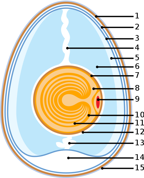
English:
Diagram of a chicken egg:
Español:
Morfología de huevo de gallina:
Italiano:
Morfologia dell'uovo di pollo:
Galego:
Diagrama dun ovo de galiña:
العربية: أجزاء بيض الدجاج
|
| Date | |
| Source | graphic created by de:Benutzer:Horst Frank, SVG version by cs:User:-xfi- |
| Author | de:Benutzer:Horst Frank, SVG code cs:User:-xfi- |
| Permission (Reusing this file) |
Horst Frank released it under GFDL |
| Other versions | Image:Ei1.jpg ; corrected : Image:Anatomy of an amiotic egg.svg ; image:Anatomy of an egg Ar.svg |

|
Permission is granted to copy, distribute and/or modify this document under the terms of the GNU Free Documentation License, Version 1.2 or any later version published by the Free Software Foundation; with no Invariant Sections, no Front-Cover Texts, and no Back-Cover Texts. A copy of the license is included in the section entitled GNU Free Documentation License. |
| This file is licensed under the Creative Commons Attribution-Share Alike 3.0 Unported license. | ||
| ||
| This licensing tag was added to this file as part of the GFDL licensing update. |
cs:Image:Anatomy of an egg.svg
File history
Click on a date/time to view the file as it appeared at that time.
| Date/Time | Thumbnail | Dimensions | User | Comment | |
|---|---|---|---|---|---|
| current | 12:53, 15 June 2021 | | 566 × 702 (9 KB) | MdsShakil | File uploaded using svgtranslate tool (https://svgtranslate.toolforge.org/). Added translation for bn. |
| 01:08, 8 August 2006 |  | 566 × 702 (6 KB) | -xfi- |
File usage
The following page uses this file:
Global file usage
The following other wikis use this file:
- Usage on ar.wikipedia.org
- Usage on bg.wikipedia.org
- Usage on bn.wikipedia.org
- Usage on cs.wikipedia.org
- Usage on da.wikipedia.org
- Usage on de.wikipedia.org
- Usage on el.wikipedia.org
- Usage on en.wikipedia.org
- Usage on en.wiktionary.org
- Usage on eo.wikipedia.org
- Usage on et.wikipedia.org
- Usage on eu.wikipedia.org
- Usage on fa.wikipedia.org
- Usage on fi.wikipedia.org
- Usage on fr.wikipedia.org
- Usage on fr.wiktionary.org
- Usage on hr.wiktionary.org
- Usage on hu.wikipedia.org
- Usage on hu.wikibooks.org
- Usage on hy.wikipedia.org
- Usage on incubator.wikimedia.org
- Usage on it.wiktionary.org
- Usage on ja.wikipedia.org
- Usage on ko.wikipedia.org
- Usage on ku.wiktionary.org
- Usage on ky.wikipedia.org
- Usage on lfn.wikipedia.org
- Usage on lt.wikipedia.org
View more global usage of this file.