Comparative anatomy

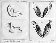
Comparative anatomy is the scientific comparison of animal bodies. The purpose of comparative anatomy is to see their working structure, and to decide upon the phylogenetic relationships between different groups of animals. The separation of animals into phyla is done mainly by comparative anatomy: see List of animal phyla.
The main techniques used are dissection and microscopy. Dissection is an ancient method used to find out the inside structure of a living thing (usually done after it is dead). It is still used by medical students to learn about the details of the human body. Simple microscopes were first invented in the 17th century, and the compound microscope (still used today) became available in the 19th century. The purpose of microscopy is to let us see the small details of structure. Also, the careful comparison of large collections of animals (usually in museums) is often done.
The great era of comparative anatomy was from about 1800 to about 1950. It was used by those who did not believe in evolution, such as Georges Cuvier, and by those who did, such as Thomas Henry Huxley. Charles Darwin himself used comparative anatomy as the main tool in his work on barnacles. Nowadays, the main method used to find out relationships is molecular evolution, which uses DNA sequence analysis. However, for many research purposes, zoologists still dissect animals. To get a degree in biology, you need to know about the structure of animals (and plants).
