
File:Eye I.jpg

Size of this preview: 614 × 599 pixels. Other resolutions: 246 × 240 pixels | 492 × 480 pixels | 787 × 768 pixels | 1,049 × 1,024 pixels | 2,098 × 2,048 pixels | 5,000 × 4,880 pixels.
Original file (5,000 × 4,880 pixels, file size: 17.81 MB, MIME type: image/jpeg)
| This is a file from the Wikimedia Commons. Information from its description page there is shown below. Commons is a freely licensed media file repository. You can help. |
| Description |
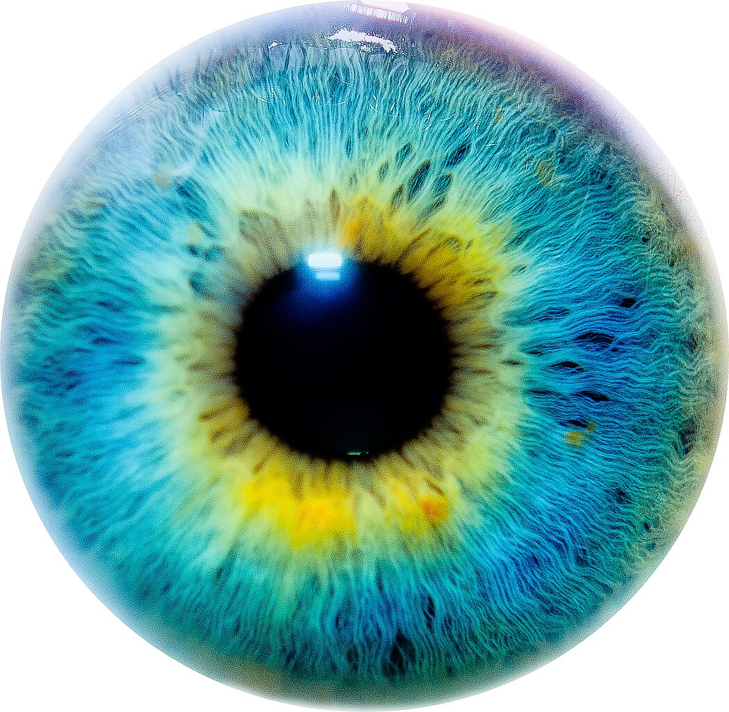
As seen by me. Facebook Fan Page | Twitter |
||
| Date | |||
| Source | Flickr: Eye I | ||
| Author | Thomas Tolkien | ||
| Permission (Reusing this file) |
This file is licensed under the Creative Commons Attribution 2.0 Generic license.
|
File history
Click on a date/time to view the file as it appeared at that time.
| Date/Time | Thumbnail | Dimensions | User | Comment | |
|---|---|---|---|---|---|
| current | 00:03, 21 November 2020 | | 5,000 × 4,880 (17.81 MB) | SteinsplitterBot | Bot: Image rotated by 180° |
| 18:00, 4 August 2017 |  | 5,000 × 4,880 (17.81 MB) | SteinsplitterBot | Bot: Image rotated by 180° | |
| 23:13, 9 August 2011 |  | 5,000 × 4,880 (17.28 MB) | Flickr upload bot | Uploaded from http://flickr.com/photo/38585972@N04/5718897981 using Flickr upload bot |
File usage
The following page uses this file:
Global file usage
The following other wikis use this file:
- Usage on ba.wikipedia.org
- Usage on en.wikipedia.org
- Actinic conjunctivitis
- Fluocinolone acetonide
- Band keratopathy
- Streff syndrome
- Eye testing using speckle
- E chart
- Ardalan–Shoja–Kiuru syndrome
- Molecular Vision
- Mizuo–Nakamura phenomenon
- Acute zonal occult outer retinopathy
- Noor Eye Hospital
- Floppy eyelid syndrome
- Foveal avascular zone
- Template:Ophthalmology-stub
- Journal of Pediatric Ophthalmology and Strabismus
- John Jeffries II
- Meibography
- Evaporimetry
- Wilmer Ophthalmological Institute
- Infracyanine green
- Anterior chamber paracentesis
- Transcorneal electrical stimulation
- Usage on fr.wikipedia.org
- Usage on lmo.wikipedia.org
- Usage on vi.wikipedia.org
- Usage on www.wikidata.org