Ribosome
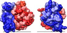
Ribosomes are important cell organelles. They are micromolecular machines that make proteins. They do this through RNA translation, building proteins from amino acids using messenger RNA as a template. Ribosomes are found in all living cells, prokaryotes as well as eukaryotes.
Ribosomes are made of two parts: the large and small subunits. Each subunit is made of a mixture of protein and rRNA that are made in the nucleolus of a cell. After being made, ribosomes move from the nuclear envelope to the cytoplasm. Most ribosomes sit on the endoplasmic reticulum, but are also found throughout the cytoplasm.
Human cells can have up to 10 million ribosomes in every cell. In order to create each ribosome, cells have many copies of rRNA genes. In humans, about 400 rRNA genes are inherited across five chromosomes.[1][2]


Structure
[change | change source]
Ribosomes are made out of two things: a small ribosomal subunit that reads the mRNA, while the large subunit joins amino acids to form a polypeptide chain. Each subunit is composed of one or more ribosomal RNA (rRNA) molecules and a variety of proteins.
Eukaryotes have 80S ribosomes, each consisting of a small (40S) and large (60S) subunit.[3][4][5] Their small subunit has a 16S RNA sub-unit (consisting of 1540 nucleotides) bound to 21 proteins. The large subunit has a 5S RNA (120 nucleotides), a 28S RNA (4700 nucleotides), a 5.8S RNA (160 nucleotides) subunits and 46 proteins.[4][6][7]
Types of ribosome
[change | change source]Ribosomes evolved as cells evolved. Prokaryote (bacterial) ribosomes have just a single RNA chain. Archaeal ribosomes are similar.
Only Eukaryotic ribosomes have the full gear, which is a total 80S ribosome with a small 40S unit plus a large 60S subunit.
References
[change | change source]- ↑ Carey, Nessa 2015. Junk DNA: A journey through the dark matter of the genome, p149. Icon Books ISBN 978-1-84831-826-7
- ↑ Zentner G.E. et al July 2011. Nucleic Acid Research 39(12) 4949–4960.
- ↑ The unit of measurement is the Svedberg unit, a measure of the rate of sedimentation in centrifugation rather than size. This accounts for why fragment numbers do not add up (80S is made of 40S and 60S).
- ↑ 4.0 4.1 Ben-Shem A. et al 2011 (2011). "The structure of the eukaryotic ribosome at 3.0 Å resolution". Science. 334 (6062): 1524–1529. Bibcode:2011Sci...334.1524B. doi:10.1126/science.1212642. PMID 22096102. S2CID 9099683.
{{cite journal}}: CS1 maint: numeric names: authors list (link) - ↑ Rabl et al 2010 (2011). "Crystal structure of the eukaryotic 40S ribosomal subunit in complex with initiation factor 1". Science. 331 (6018): 730–736. Bibcode:2011Sci...331..730R. doi:10.1126/science.1198308. hdl:20.500.11850/153130. PMID 21205638. S2CID 24771575.
{{cite journal}}: CS1 maint: numeric names: authors list (link) - ↑ Alberts, Bruce et al 2002. The molecular biology of the cell. 4th ed, Garland Science, 342. ISBN 0-8153-3218-1
- ↑ Klinge et al 2011 (2011). "Crystal structure of the eukaryotic 60S ribosomal subunit in complex with initiation factor 6". Science. 334 (6058): 941–948. Bibcode:2011Sci...334..941K. doi:10.1126/science.1211204. PMID 22052974. S2CID 206536444.
{{cite journal}}: CS1 maint: numeric names: authors list (link)
