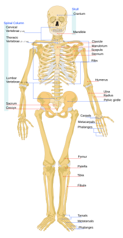
Human skeleton

human skeleton is the internal framework of the body. It is made up of about 300 bones at birth. As some bones join together, there are 206 bones in adulthood.[1] The bones are at their strongest around age 20. The human skeleton can be divided into the axial skeleton and the appendicular skeleton. The axial skeleton is formed by the vertebral column, the rib cage, the skull and other associated bones. The appendicular skeleton, which is attached to the axial skeleton, is formed by the shoulder girdle, the pelvic girdle and the bones of the upper and lower limbs.
The human skeleton performs six major functions. These are: support, movement, protection, production of blood cells, storage of minerals, and endocrine regulation.
The male and female skeletons are not so different as those of many other primates. There are subtle differences between sexes in the morphology of the skull, teeth, long bones, and pelvis. In general, female skeletal elements tend to be smaller and less robust than corresponding male elements.
The human female pelvis is different from that of males in order to facilitate child-birth.[2] The hips on a female are proportionately wider than males, and so the ball joints at the top of the legs are set more apart than in males. This, and the pelvic shape, gives a birth channel which allows the newborn foetus to pass through. The critical factor is the baby's head, which is much larger than in other primates.
Unlike most primates, human males do not have penile bones.[3] This is an adaptation to the human upright stance.
References[change | change source]
- ↑ Mammal anatomy: an illustrated guide. New York: Marshall Cavendish. 2010. p. 129. ISBN 978-0-7614-7882-9.
- ↑ Thieme Atlas of Anatomy, 2006, p 113.
- ↑ Ford, Clellan Stearns; Beach, Frank Ambrose (1980). Patterns of Sexual Behavior. Greenwood Publishing Group. ISBN 978-0-313-22355-6.
