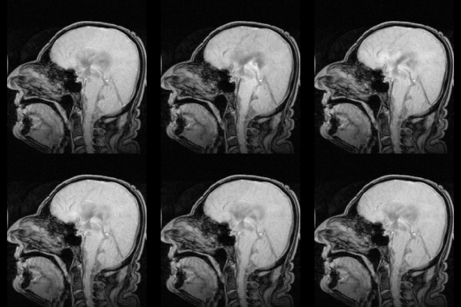
Cerebrospinal fluid


Cerebrospinal fluid (CSF) bathes and protects the central nervous system (the brain and the spinal cord). "Cerebro" means "brain"; "spinal" is a short version of "spinal cord"; and fluid is a liquid.
CSF is made by networks of blood vessels called choriod plexuses in each of the brain's four ventricles.[1]
CSF flows through the subarachnoid space – the space between the two layers of meninges that are closest to the brain (the arachnoid layer and the pia mater). CSF also fills the brain's ventricles, and flows down the middle of the spinal cord (the central canal).[2]
Contents[change | change source]
CSF is created from blood plasma (the liquid part of blood), so CSF's contents are very similar to plasma's.[3] CSF is 99% water. It carries:[3]
- Protein (though much less than plasma carries)
- Glucose (sugar)
- Nutrients
- Neurotransmitters (chemical messengers)
- Electrolytes (salts), like sodium, potassium, calcium, magnesium, and chloride.
There should be very few white blood cells in the CSF, or none at all. There should be no red blood cells in the CSF.[4]
What CSF does to the brain and the surrounding components[change | change source]
CSF helps the brain float[change | change source]
Being surrounded by CSF helps the brain float inside the skull, like a buoy in water. Because the brain is surrounded by fluid, it floats like it weighs only 2% of what it really does.[5] If the brain did not have CSF to float in, it would sit on the bottom of the skull. The brain's weight would push the bottom of the brain against the skull. Blood would not be able to get to the bottom of the brain because the blood vessels would be getting crushed by the weight of the brain on top of them. Without getting blood (and the oxygen it carries), the neurons in the bottom of the brain would die.[6]
CSF is the brain's cushion[change | change source]
CSF protects the brain by acting like a cushion. Without CSF, every time a person moved their head, their brain would hit the inside of their skull. This could injure the brain.
When a person hits their head, CSF acts like the airbag in a car and can sometimes keep the brain from slamming into the inside of the skull. However, when a person hits their head very hard – in a car accident, for example – the CSF cannot protect the brain from hitting the skull. This can cause concussions, bleeding in the brain, brain damage, or even death.[6]

CSF rinses toxins out of the brain[change | change source]
The brain's cells do chemical reactions to change one chemical to another chemical that the brain needs.[7] Sometimes, after a chemical reaction, chemicals that the brain does not need are left over. These chemicals are called "waste products." For example, when the brain's cells use oxygen and glucose (sugar) to create energy, carbon dioxide (CO2) is left over.[7] Too much carbon dioxide can poison the brain.[2]
To keep waste products from building up in the CSF, the choroid plexuses make new CSF about four times a day.[1] The old CSF drains out into the bloodstream, bringing waste products and toxins with it. In this way, the CSF rinses things that could hurt the brain out into the bloodstream.[8] The bloodstream can then carry these chemicals to organs that can get rid of them, like the kidneys and the lungs.[9] For example, the bloodstream carries carbon dioxide to the lungs, where it can be breathed out.
Testing CSF[change | change source]
Doctors can use samples of CSF to find out if a person has a brain infection, like meningitis, encephalitis, or syphilis. CSF samples can also show bleeding from certain parts of the brain. Swelling in the brain, caused by some inflammatory diseases like multiple sclerosis, can show in a CSF sample as well.[10][4]
Usually, doctors take samples of CSF by doing a lumbar puncture (spinal tap).[4]
Results[change | change source]
Normal CSF should be clear and colorless, with no red blood cells, and very few white blood cells.[4]
Signs of a brain infection include:[10]
- CSF that is cloudy, yellow, or pink
- More protein in the CSF than normal
- More white blood cells in the CSF than normal
- Less glucose (sugar) in the CSF than normal
- Bacteria, viruses, fungi, or other pathogens in the CSF
Protein levels that are higher than normal can be a sign of inflammation in the brain. However, they can also be a sign of other problems, like a bleed in the brain; a brain tumor; epilepsy; and "acute alcoholism."[11]
Cancer cells in the CSF are a sign that a person has brain cancer, or has cancer that started somewhere else and spread to the brain.[12]
Problems with CSF[change | change source]
Too much[change | change source]
Head injuries and some diseases can make too much CSF build up in the brain.[13] This is called hydrocephalus. The extra fluid puts pressure on (squeezes) the brain. This is called increased intracranial pressure. If the pressure gets high enough, the brain's blood vessels get crushed, and blood cannot get to the brain. If a person's brain does not get enough blood and oxygen, they will become unconscious and their brain cells will die. Eventually, the person will die.[2]
Without treatment, 6 out of every 10 people with hydrocephalus will die.[14] The rest will have physical disabilities, intellectual disabilities, and/or other brain problems.[14]
Not enough[change | change source]

Some problems can cause CSF to leak out through a hole or tear in the dura mater (the layer of the meninges that is next to the skull and the bones of the spinal cord). CSF leaks are usually a complication of a lumbar puncture or certain kinds of surgery on the head, neck, or brain. However, an injury to the head or spine can also cause a CSF leak. Sometimes, doctors cannot find a cause for a CSF leak.[15]
Most people with CSF leaks get better on their own, after the hole in the dura mater heals.[15] However, some CSF leaks can cause serious complications:[16]
- Bacteria, viruses, or other pathogens can get in through the same hole CSF is leaking out of. This can cause brain infections like meningitis.
- Without enough CSF to float in, the brain sits on the bottom of the skull. The brain's weight can:
- Crush the blood vessels in the bottom of the brain
- Press on the brainstem, causing very low blood pressure
- Push the brain down through the hole in the bottom of the skull, where the brainstem turns into the spinal cord. This can cause paralysis, brain damage, coma, and death.
Related pages[change | change source]
References[change | change source]
- ↑ 1.0 1.1 Chuder, Eric H. "The Ventricular System and CSF (Cerebrospinal Fluid)". faculty.washington.edu. National Center for Research Resources.
{{cite web}}: Missing or empty|url=(help) - ↑ 2.0 2.1 2.2 Mostovich, Joseph J.; Hafen, Brent Q.; Karren, Keith J. (October 28, 2009). Prehospital Emergency Care (9th ed.). Prentice Hall. ISBN 9780135028100.
- ↑ 3.0 3.1 Wood, James H. (June 29, 2013). Neurobiology of Cerebrospinal Fluid 2. Springer Science and Business Media. p. 3. ISBN 9781461592693.
- ↑ 4.0 4.1 4.2 4.3 Kantor, David (June 1, 2015). "CSF Cell Count". MedlinePlus. United States National Library of Medicine.
{{cite web}}: Missing or empty|url=(help) - ↑ Noback, Charles; Strominger, Norman L.; Demarest, Robert J.; Ruggiero, David A. (2005). The Human Nervous System. Humana Press. p. 93. ISBN 978-1-58829-040-3.
- ↑ 6.0 6.1 Saladin, Kenneth (2007). Anatomy and Physiology: The Unity of Form and Function. McGraw Hill. p. 520. ISBN 978-0-07-287506-5.
- ↑ 7.0 7.1 Schousboe A; Walls AB; et al. 2015 (2015). "Astroglia and Brain Metabolism: Focus on Energy and Neurotransmitter Amino Acid Homeostasis". Colloqium Series on Neuroglia in Biology and Medicine: From Physiology to Disease. 2 (4). Morgan & Claypool Publishers: 1–64. doi:10.4199/C00130ED1V01Y201506NGL007. Archived from the original on June 15, 2022. Retrieved February 6, 2016.
{{cite journal}}: CS1 maint: multiple names: authors list (link) CS1 maint: numeric names: authors list (link) - ↑ Iliff JJ; Wang M; et al. 2012 (August 2012). "A paravascular pathway facilitates CSF flow through the brain parenchyma and the clearance of interstitial solutes, including amyloid β". Science Translational Medicine. 4 (147): 147ra111. doi:10.1126/scitranslmed.3003748. PMC 3551275. PMID 22896675.
{{cite journal}}: CS1 maint: multiple names: authors list (link) CS1 maint: numeric names: authors list (link) - ↑ Ropper, Allan H.; Brown, Robert H. (March 29, 2005). Adams and Victor's Principles of Neurology (8th ed.). McGraw-Hill Professional. p. 530. ISBN 978-0071416207.
- ↑ 10.0 10.1 "Lumbar Puncture (Spinal Tap)". The Mayo Clinic. Mayo Foundation for Medical Education and Research. December 6, 2014. Retrieved February 6, 2016.
- ↑ Seehusen DA; Reeves MM; et al. 2003 (2003). "Cerebrospinal fluid analysis". American Family Physician. 68 (6): 1103–8. PMID 14524396. Retrieved February 6, 2016.
{{cite journal}}: CS1 maint: multiple names: authors list (link) CS1 maint: numeric names: authors list (link) - ↑ Weston CL; Glantz MJ; et al. 2011 (2011). "Detection of cancer cells in the cerebrospinal fluid: Current methods and future directions". Fluids and Barriers of the CNS. 8 (14). BioMed Central: 14. doi:10.1186/2045-8118-8-14. PMC 3059292. PMID 21371327.
{{cite journal}}: CS1 maint: multiple names: authors list (link) CS1 maint: numeric names: authors list (link) - ↑ "Hydrocephalus Fact Sheet". National Institute of Neurological Disorders and Stroke. United States National Institutes of Health. February 3, 2016. Archived from the original on July 27, 2016. Retrieved February 6, 2016.
- ↑ 14.0 14.1 Kaneshiro, Neil K. (December 4, 2013). "Hydrocephalus". MedlinePlus. United States National Library of Medicine, National Institutes of Health. Retrieved February 6, 2016.
- ↑ 15.0 15.1 Campellone, Joseph V. (July 27, 2014). "CSF Leak". MedlinePlus. United States National Library of Medicine, National Institutes of Health. Retrieved February 6, 2016.
- ↑ Schievink WI 2006 (2006). "Spontaneous Spinal Cerebrospinal Fluid Leaks and Intracranial Hypotension". Journal of the American Medical Association. 295 (19): 2286–96. doi:10.1001/jama.295.19.2286. PMID 16705110.
{{cite journal}}: CS1 maint: numeric names: authors list (link)
